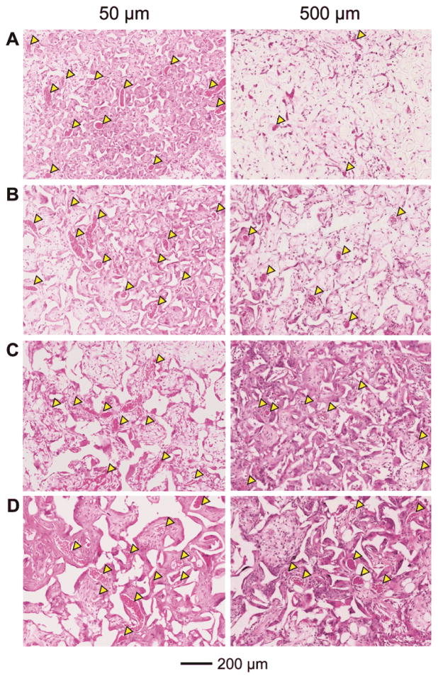
Figure 6.
Representative hematoxylin and eosin stained tissue sectionsof subcutaneously implanted scaffolds with pore sizes of (A) 79, (B) 147, (C) 224, and (D) 312 μm, respectively, at 4 weeks post implantation at approximately 50 μm (left column) and 500 μm (right column) in depth from the top surface of the scaffolds. Blood vessels are indicated by yellow arrowheads.

