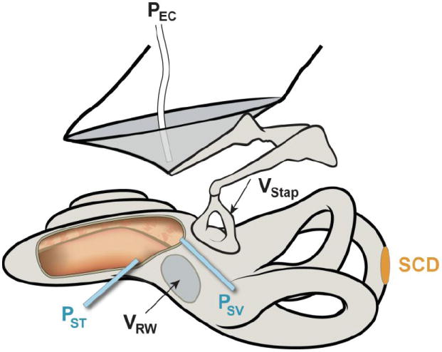
Figure 1.

Illustration of a left ear demonstrating a superior semicircular canal dehiscence (SCD) along with measurements made. Measurements included sound pressure in the ear canal (PEC) with a probe-tube microphone, stapes velocity (VStap) and round-window velocity (VRW) with laser Doppler vibrometry and sound pressures in scala vestibuli (PSV) and scala tympani (PST) measured simultaneously with micro-optical pressure transducers. For illustration purposes only, the scalae are shown opened (cut-out area to the left of the round window) to show the placement of the transducers within the two perilymphatic scalae.
