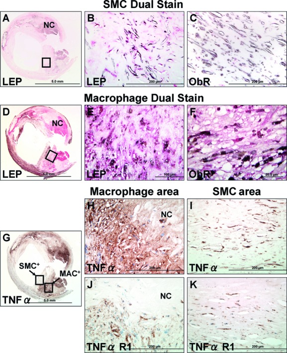
Figure 2.

Colocalization of SMCs and macrophages with leptin and its receptor (ObR) and with tumor necrosis factor-α (TNF-α) and its receptor (TNF-αR1) in a human carotid fibroatheroma. A, Dual staining for SMCs (brown-black) and leptin (LEP, red). B, SMCs near the fibrous cap represented by the area within the black box in (A) are positive for leptin. C, SMCs weakly positive for leptin receptors (ObR). D, Dual staining for macrophages (brown-black) and leptin (LEP, red). E and F, Region represented by the area within the black box in (D); macrophages are positive for both leptin and ObR. G, immunostain for TNF-α. H to K, Images within the black boxes in (G) correspond to SMC and macrophage-rich areas (MAC), respectively. TNF-α and its receptor (TNF-αR1) are expressed in both regions. SMCs indicates smooth muscle cells.
