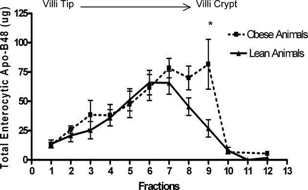
Figure 4.

Cell-associated apoB48 mass along the length of the intestinal villus. Approximately 30 cm of jejunal tissue was excised and used for enterocyte isolation by the Weiser method (see details in Methods). ApoB48 mass was determined for each isolated cell fraction via immunoblotting. MetS indicates metabolic syndrome. *P<0.05 represents statistical significance in apoB48 from isolated cell fractions between phenotypically lean (n=8) and MetS (insulin-resistant) (n=8) rats.
