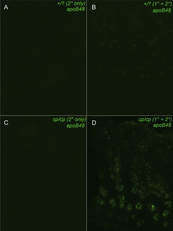
Figure 5.

Immunohistochemical visual of the distribution of ApoB48 along the intestinal villus. A and B, Intestinal sections from lean animals (+/?) using secondary antibody alone or primary and secondary antibody, respectively. C and D, Sections from a MetS (cp/cp) rat treated with secondary antibody alone or primary and secondary antibody, respectively. MetS indicates metabolic syndrome. Images were captured at 20× magnification.
