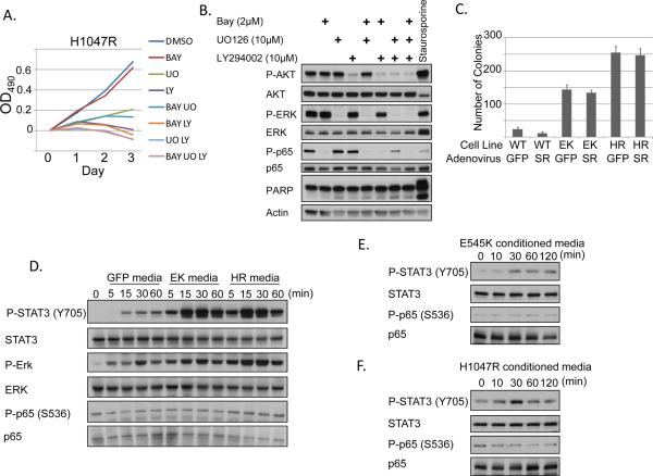
Figure 5. Secreted factors from cells expressing oncogenic PI3K mutations activate STAT3.
A) GF-deprived MCF10A cells expressing PIK3CA H1047R were treated for 3 days with the indicated inhibitors. Media and inhibitors were replaced daily. BAY = 2μM BAY-65-1942. LY = 10μM LY294002. UO = 10μM UO126 and cells were evaluated by MTT assay. B) Growth factor-starved MCF10A cells expressing H1047R were treated for 2 days with the indicated inhibitors. Media and inhibitors were replaced daily. Lysates were evaluated for PARP cleavage. 1μM staurosporine (positive control) was added 6h prior to lysis. C) MCF10A cells expressing PIK3CA WT, E545K, or H1047R were infected with adenovirus expressing either GFP or IκBα superrepressor (SR) and plated in 0.6% Bacto agar in MCF10A media lacking EGF and insulin. Colonies were counted after 25 days. D) MCF10A cells expressing GFP, PIK3CA E545K, or PIK3CA H1047R were starved for 24h. Conditioned media from these cells was used to treat THP-1 monocytes for 0–60 minutes. Lysates were evaluated by immunoblot. E–F) MCF10A cells stably expressing PIK3CA E545K (E) or H1047R (F) were starved for 24h. Conditioned media from these cells was then used to stimulate starved parental MCF10A cells for 0–120 minutes. Lysates were evaluated by immunoblot.

