Figure 5.

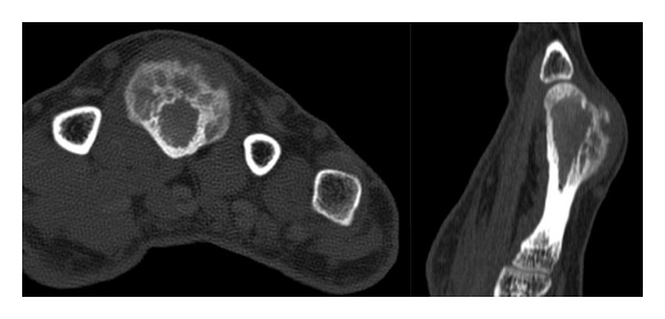
CT axial and sagittal images of the third metacarpal demonstrating a small focal area of continuity between the medullary cavity of the lesion and that of the underlying bone.

CT axial and sagittal images of the third metacarpal demonstrating a small focal area of continuity between the medullary cavity of the lesion and that of the underlying bone.