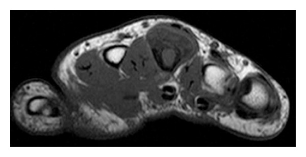
Figure 7.

On an axial T1-weighted preoperative MR image of the area, the lesion is composed predominantly of central signal isointense to that of the normal bone marrow with a thin low signal periphery consistent with the cortical bone. Direct continuity with the underlying marrow of the bone is not noted.
