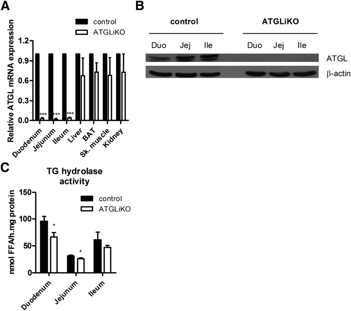
Fig. 1.
ATGL is knocked out specifically in the small intestine. A: ATGL mRNA was drastically down-regulated in all three parts of the small intestine (duodenum, jejunum, ileum) but unchanged in control tissues (liver, brown adipose tissue, skeletal muscle, kidney). Data represent mean values ± SEM (n = 3). ***P ≤ 0.001. :) Protein lysates of pools from three mice of each genotype were separated by SDS-PAGE. ATGL protein expression was analyzed by Western blotting. The expression of β-actin served as loading control. C: TG hydrolase activity was determined in duodenum, jejunum, and ileum of HFD-fed mice. Data represent mean values ± SEM (n = 4). *P ≤ 0.05.

