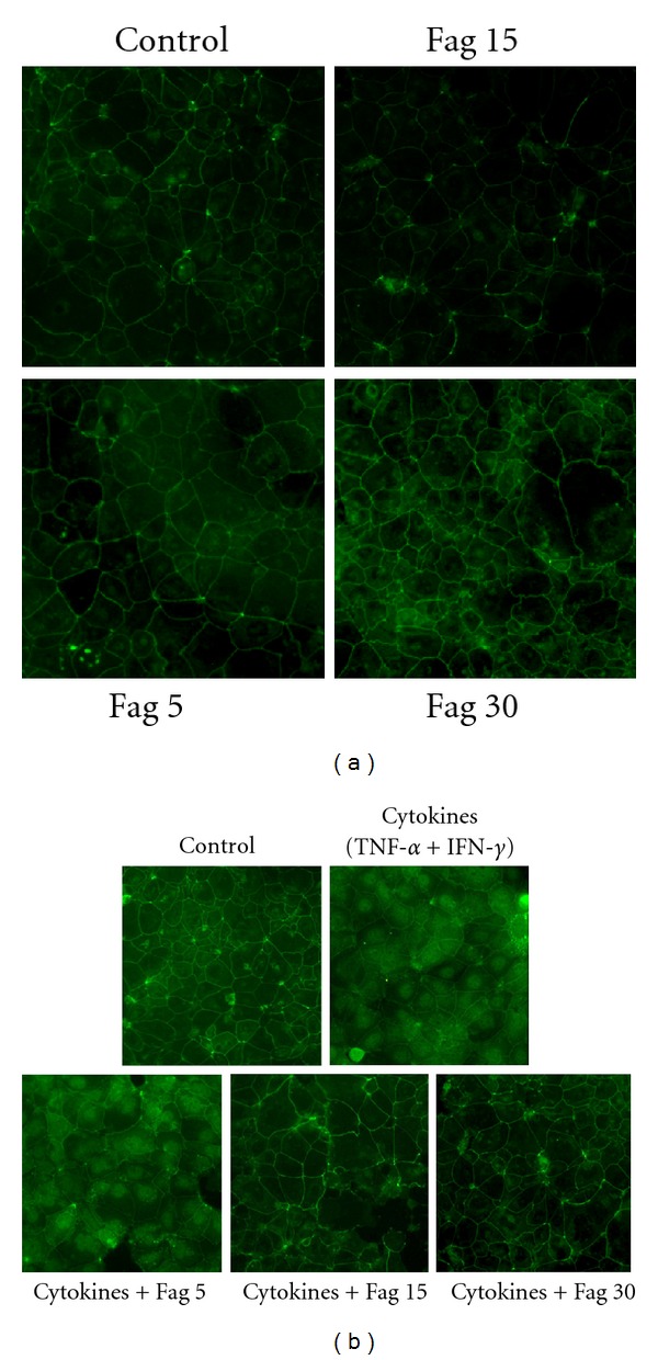
Figure 8.

Immunofluorescence of claudin-1 in Caco-2 cell monolayers. (a) The cells were incubated with Fag (0, 5, 15, 30 μg/mL) and stained by claudin-1 for 24 h. (b) The cells were incubated with Fag (0, 5, 15, 30 μg/mL) and cytokines (100 ng/mL TNF-α and 100 ng/mL IFN-γ) or Fag alone for 24 h. The images were collected by LSCM (×200).
