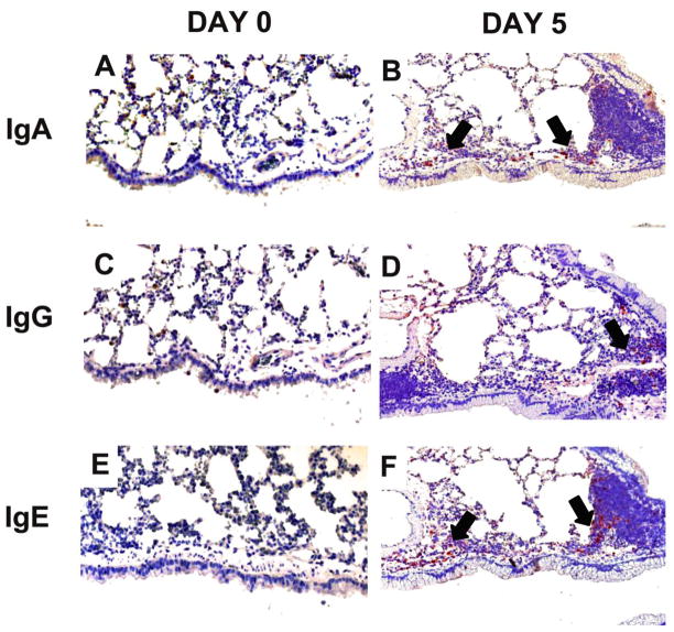
Figure 5. Effect of inhalation of A. fumigatus conidia on IgA, IgG, and IgE antibody producing cells in the allergic mouse lung.
Mice were exposed to A. fumigatus according to the schedule shown in figure 1. Immunohistochemical staining was used to identify immunoglobulin producing cells in the lung tissue sections of day 0 (sensitized but not challenged with A. fumigatus) and day 5 allergic animals. IgA, IgG and IgE producing cells increased after allergen challenge with maximum numbers at day 5. (A, C, & E) Naïve BALB/c lungs had very few cells that localized Ig (IgA, IgG, and IgE). (B, D, & F) Day 5 BALB/c lungs contained substantial numbers of IgA, IgG, and IgE positive cells localized around the airways (indicated by arrows).

