Fig. 2.
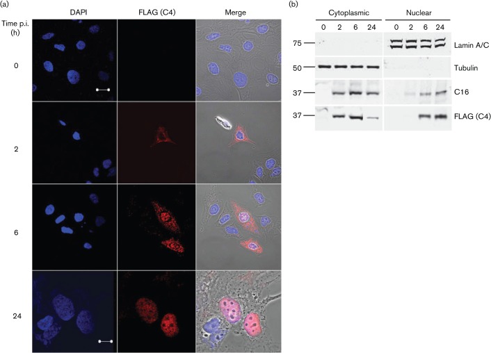
Subcellular localization of C4. (a) Immunofluorescence. HeLa cells were infected with vC4-TAP at 0.5 p.f.u. per cell for the indicated times. Cells were washed with PBS, fixed and stained with anti-FLAG mAb. The localization of C4/FLAG (red; middle panels), DNA stained with DAPI (blue; left panels) and phase-contrast/merged images (right panels) are shown. Bars, 20 µm (0–6 h); 10 µm (24 h). (b) Immunoblotting. BSC-1 cells were infected at 10 p.f.u. per cell with vC4-TAP for the indicated times, harvested and fractionated into cytoplasmic and nuclear fractions. Protein fractions were resolved by SDS-PAGE and analysed by immunoblotting with antibodies against the indicated proteins. The positions of molecular mass markers are shown (kDa).

