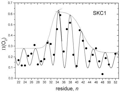
Figure 3.
Oxygen accessibilities, Π(O2), of site-directed spin labels attached to the first transmembrane segment of the SKC1 K+-channel. Experimental values (29) are given by ●, as a function of the labeled residue position in systematic cysteine-substitution mutants. Dashed lines give the envelope of the oxygen permeation profile at the position of the lipid-facing residues, according to Eq. 2. Solid lines are the complete oxygen concentration profiles, modulated by the helical residue periodicity of exposure, according to Eq. 6. The two halves of the membrane, residues 22–36 and 36–50, respectively, are fitted separately as described in the text.

