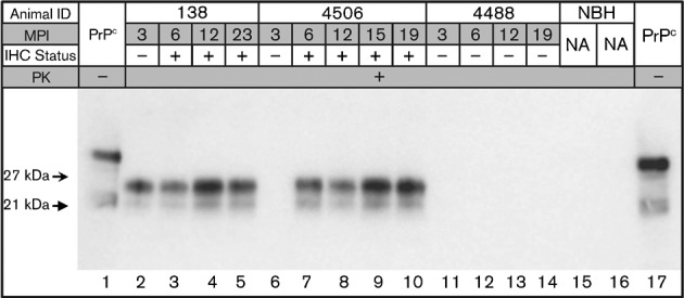
Fig. 1.

Western blot analysis of tonsil biopsy sPMCA from deer numbers 138 (inoculated intracranially with CWD-positive brain tissue), 4506 (inoculated intravenously with CWD-positive white blood cells) and 4488 (inoculated intracranially and per os with CWD-negative brain tissue) (Table 1). Biopsy dates, in months post-inoculation (MPI), as well as IHC results, are given for each individual and tonsil biopsy, respectively. Non-proteinase K-treated brain homogenate is included in lanes 1 and 17 for band size reference, and unspiked normal brain homogenate (NBH) controls are shown in lanes 15 and 16. na, Not applicable.
