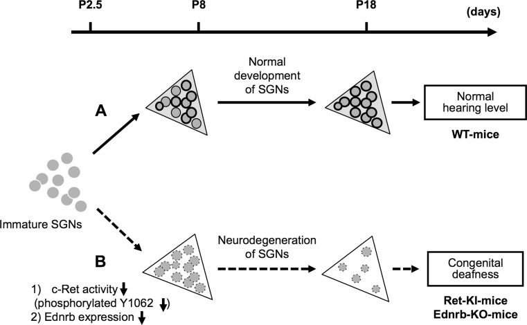
Fig. 1.
Schematic summary of congenital deafness caused by neurodegeneration of spiral ganglion neurons (SGNs) in c-Ret-knock-in-mice and Ednrb-knock-out-mice. The x-axis indicates age (days after birth) of mice. Triangles Rosenthal’s canals in wild-type (WT) (light gray background), or homozygous c-Ret-knock-inY1062F/Y1062F (Ret-KI) [20] and homozygous Ednrb-knock-out-mice (Ednrb-KO) (white background) [36]; gray circles/no outline immature SGNs; gray circles/thin outline SGNs; gray circles/bold outline SGNs with “phosphorylated Y1062 in c-Ret” or “expression of Ednrb”. Dark gray circles/dotted outline SGNs with “decreased phosphorylation of Y1062 in c-Ret” or “decreased expression of Ednrb”. a c-Ret-KI- and Ednrb-KO-mice suffer from congenital deafness with neurodegeneration of SGNs. b c-Ret-KIY1062F/Y1062F-mice showed no Y1062-phosphorylated SGNs even on P8, although Y1062-phosphorylated SGNs began to appear in WT mice from P8 [20]. Ednrb-KO-mice also showed undetectably low expression level of Ednrb in SGNs on P8, although Ednrb-positive SGNs began to appear in WT mice from P8 [36]

