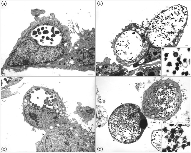
Fig. 4.
TEM of the C. trachomatis strains nvCT [Sweden2 (a) and (b)] and E/Bour [prototype reference E strain (c) and (d)] in BGMK cells. Electron micrographs were taken at 24 h [mid stage of developmental cycle (a) and (c)] and 48 h [mature inclusions (b) and (d)], and showed no major differences between the strains. The enlarged images in the lower right corner of (b) and (d) show EBs with membrane blebs.

