Fig. 2.
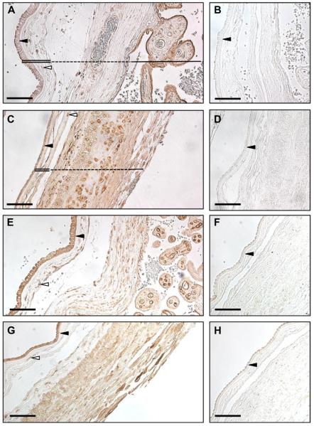
The VEGF receptors are expressed in the human fetal membranes and placenta. Immunohistochemistry was performed on formalin-fixed tissues from both the placental (A, B, E, F) and reflected (C, D, G, H) regions of the fetal membranes. The tissues sections were stained for VEGFR1 (A, C) and VEGFR2 (E, G). The respective negative controls for VEGFR1 (B, D) and VEGFR2 (F, H) are also displayed. In A and C, the amnion is indicated with a double line and the chorion with a dashed line. The solid line denotes the placenta (Pl) in A and the decidua in C. Dark arrowheads point to the amnion epithelial cell layer, and empty arrowheads identify amnion mesenchymal cells. The pictures are taken at 20× magnification and the scale bar represents 100 μm.

