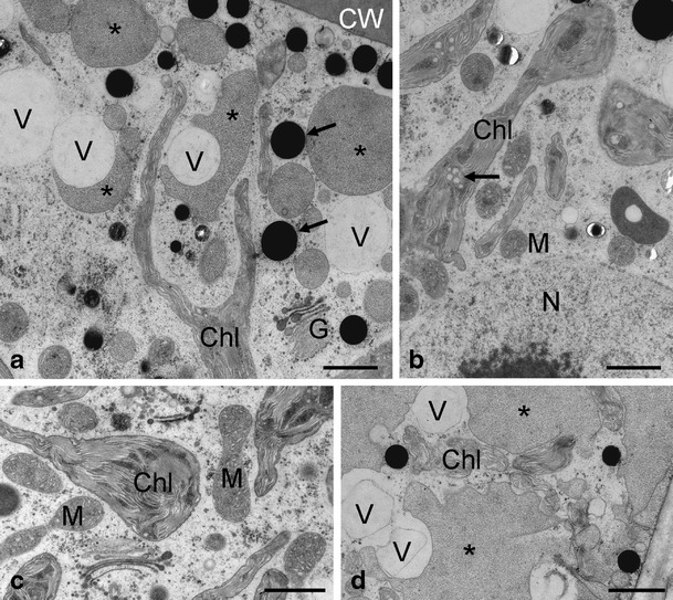
Figure 6.

Transmission electron micrographs of Zygnema sp. E after UV exposure. a Detail of the cell cortex with chloroplast lobes, substantial amount of vacuolisation, medium electron-dense compartments (asterisks) and electron dense particles (arrows); b central area, nucleus with nucleolus, mitochondria intact, the chloroplasts contain electron-dense areas and plastoglobules (arrow); c mitochondria with normal appearance, chloroplast partly swollen; d extensive medium electron-dense compartments (asterisks) in the cell cortex, vacuoles and chloroplast lobes are found in the same area. Chl chloroplast, CW cell wall, G Golgi body, L lipid body, M mitochondrion, V vacuole. Scale bars: a–d 1 μm
