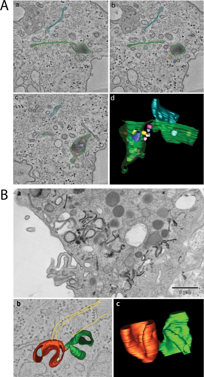
FIGURE 5:

(A) three-dimensional tomography of GA-modified endosomes. GA-treated SK-BR-3 cells were incubated with trastuzumab-HRP for 30 min on ice, prior to internalization at 37°C for 20 min in the presence of GA. The DAB reaction was performed on ice, and cells were processed for plastic embedding and electron tomography as described in Materials and Methods. Three tomographic slices at different z-axes of the original tomogram (a–c; see also Movies S1 and S2) and a three-dimensional view of the model obtained (d) are shown. Trastuzumab-HRP reaction product was found within the globular domain of the endosome, typical feature of receptors committed to degradation, as well as within the elongated tubular domain. (B) CLICs in Epon-embedded MEFs caveolin1 KO cells after 15 s of CTx-HRP internalization (+AA). (b) Dual-axis tomogram of internalized CLIC (CTx-HRP) in 300-nm-thick sections of conventionally fixed MEFs caveolin1 KO. Note the semi-ring structure with membrane invaginations and few internal vesicles (red) and the cistern domain (green; see Movie S3). Actin cytoskeleton elements (yellow) in close proximity to CLIC are depicted. (c) Three-dimensional model (Movie S4).
