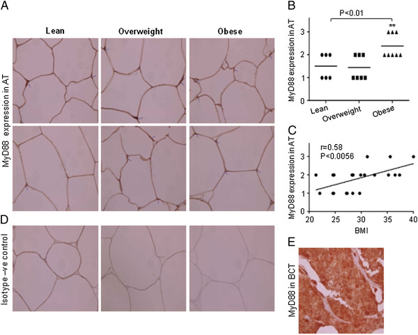
Figure 4.
Expression of MyD88 in adipose tissues. Sections of the adipose tissues from individuals (Obese 8; Overweight 7 and 6 Lean) were stained with MyD88 or with control antibodies . All sections were counterstained with hematoxylin. Positive signals appear in red (original magnification X100). Two representative sections of the adipose tissues from each group were shown. Arrowheads indicate intensity of the expression of MyD88 on the cells (A). Intensity of expression was assessed and an expression score was assigned according to following scale: 0 (no staining) and 3 (strong staining). Horizontal bars show the mean (B). Correlation and simple linear regression of AT expression of MyD88 with BMI was observed (C). Isotype –ve control (D). Section of BCT; positive control for MyD88 (E).

