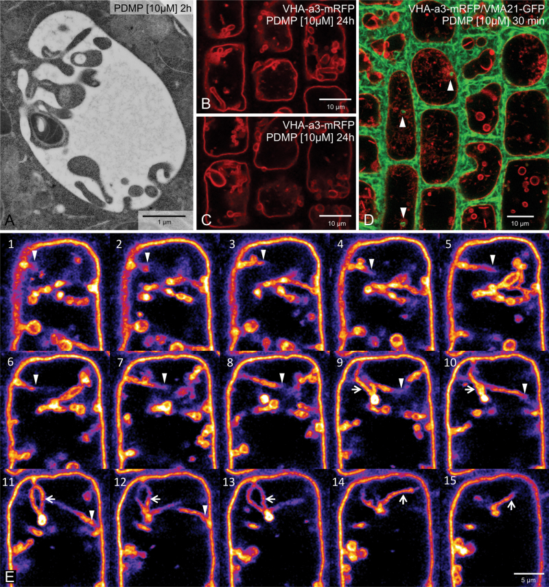
Fig. 4.
PDMP-induced vacuolar inclusions are invaginations of the tonoplast. (A) Direct connections between the tonoplast and the boundary membrane of the vacuolar inclusions can be occasionally seen in cryofixed samples. (B, C) Two optical sections from a z-stack series taken of cells in an Arabidopsis root stably expressing the tonoplast marker VHA-a3-mRFP after 10µM PDMP treatment for 24 h: tubular invaginations penetrating deep into the lumen of the vacuole are seen in several cells. (D) The endoplasmic reticulum (labelled with VMA21-mRFP, red) can be seen to penetrate into the tubular invaginations (labelled with VHA-a3-GFP, green). (E) Single optical sections of a z-stack series taken from an Arabidopis root tip cell expressing VHA-a3-mRFP: in order to emphasize the tubular invagination traversing the vacuole (arrowheads) as well as a tubular loop (arrows), the lookup table (LUT) was changed to ‘fire’ using the open source image processing software Fiji.

