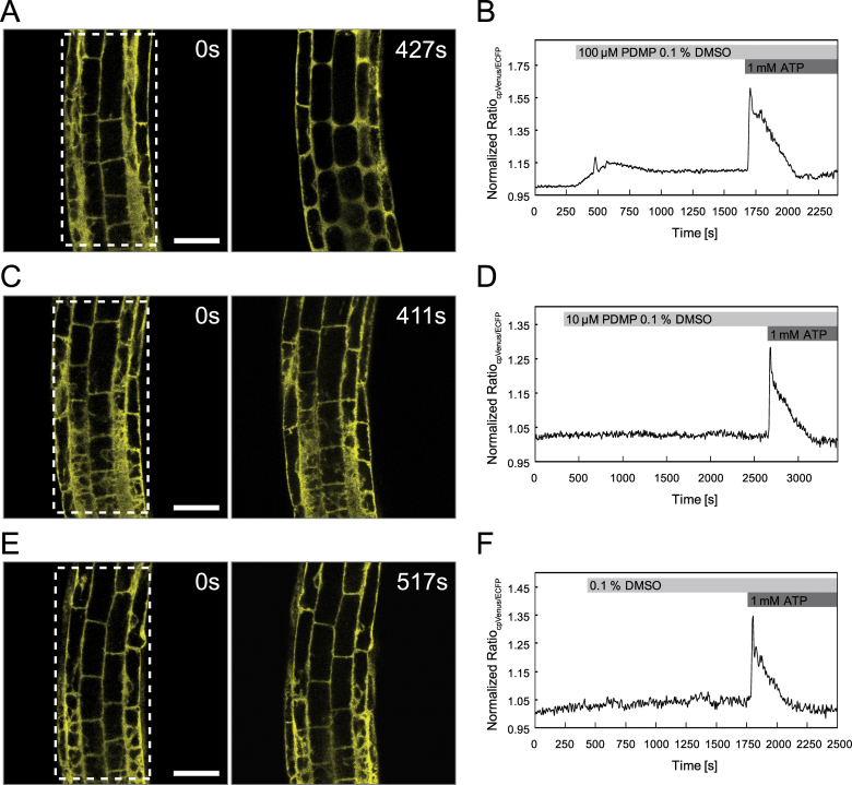
Fig. 7.
PDMP- and ATP-induced Ca2+ transients in Arabidopsis root cells. Ca2+ imaging was performed on cortical and epidermal cells of the root elongation zone of Arabidopsis seedlings expressing the Ca2+ sensor NES-YC3.6. Cytosolic Ca2+ dynamics were recorded in defined regions of interests, and the normalized ratio, indicating changes in free cytosolic [Ca2+], was monitored over time after application of different treatments. (A, C, E) Fluorescence signal cpVenus at time point zero and 100 s after PDMP/DMSO application. (B, D, F) Normalized emission ratio cpVenus/ECFP. (A, B) A moderate increase in [Ca2+]Cyt was observed after application of 100 µM PDMP, and a more pronounced Ca2+ transient was induced by treatment with 1mM ATP (B); see also Supplementary Movie S1. (C, D) In contrast to application of 1mM ATP (D), 10 µM PDMP did not induce measurable changes in [Ca2+]Cyt; see also Supplementary Movie S2. (E, F) The control treatment (0.1% DMSO) did not affect cytosolic Ca2+ levels, whereas 1mM ATP increased levels of [Ca2+]Cyt (F); see also Supplementary Movie S3. Boxes in A, C, and E indicate the regions of interest for ratiometric measurements. Data are representative of at least three experiments. Data recording was performed every 6 seconds. Bars, 50 µm.

