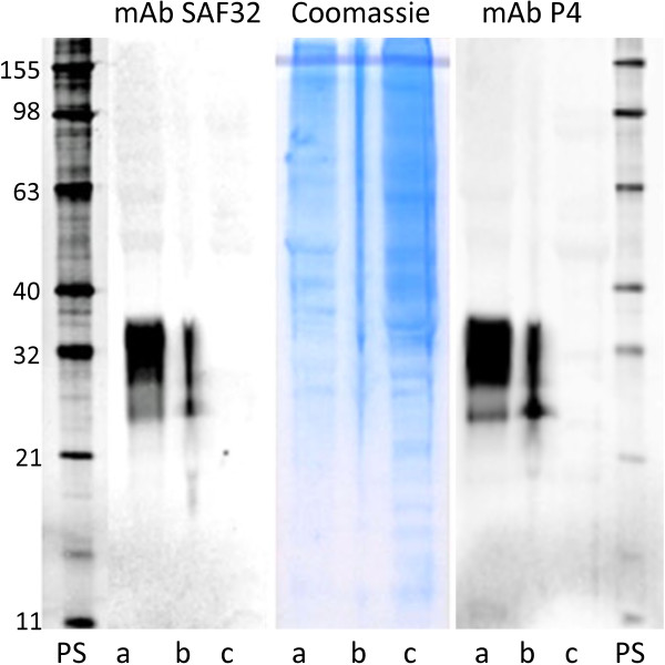
Figure 3.
Detection of PrP by Western blot. Brain samples from goats analysed using the monoclonal antibodies SAF32 (left panel) and P4 (right panel). PrP is detectable in the two wild type goats (lane a and b) but not in the homozygous 32Stop goat, (lane c). The central panel shows the presence of normal brain proteins in the three samples (Coomassie stain). PS: protein standards (kilodaltons).

