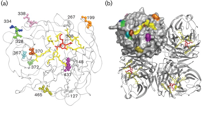
Fig. 4.
Sites identified as being positively selected, depicted on the NA globular head. Positively selected sites are shown on wire and filled-space models of the monomeric (a) and tetrameric (b) NA structure constructed using MacPyMOL on subtype N2 (PDB code 2BAT; Varghese et al., 1992). The viral receptor sialic acid, represented as red sticks, is docked into the active site, depicted as yellow sticks. All sites located in the globular head found to be positively selected are illustrated as spheres of which the residues visible on the tetrameric structure were given a colour: orange, 199; green, 328; blue, 334; magenta, 338; cyan, 367; brown, 370; lime, 372; purple, 437; olive, 465. Residues 127, 148, 267 and 308 (grey) were also found to be positively selected but were not surface exposed. Residues 43, 46 and 52 are not shown, as they are in the NA stalk domain. Numbers correlate to codon positions identified as under positive selection found within this study (Table S2).

