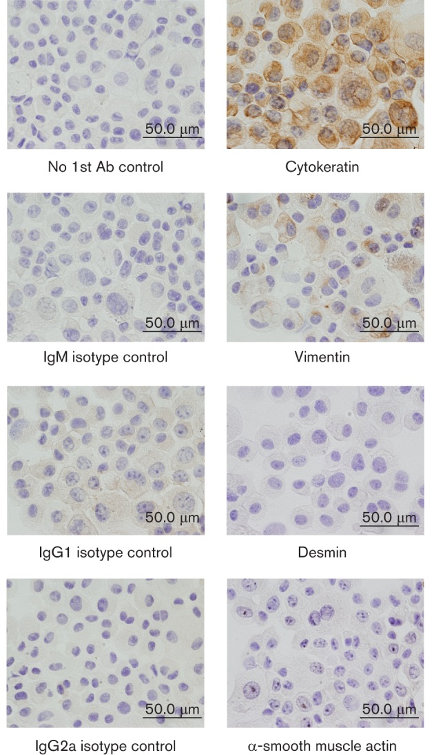
Fig. 1.

Staining of SD-PJEC cells for various epithelial, fibroblast and smooth muscle markers. Cytospins (1×10⁵ SD-PJEC cells) were fixed in acetone. The presence of cytokeratin, vimentin, alpha smooth muscle actin (ASMA) and desmin proteins was detected by immunohistochemical (IHC) staining using mAbs specific for these proteins. Staining without primary antibody but only secondary antibody of mouse IgM, IgG1 and IgG2a were used as negative and isotype controls. After washing, cells were incubated with biotinylated goat anti-mouse isotype-specific Abs. This was followed by incubation of cells with the ABC solution. Then DAB substrate was added and cells were counterstained with haematoxylin. Note, all SD-PJEC cells were positively stained for cytokeratin and few cells faintly stained for vimentin, indicating their epithelial phenotype.
