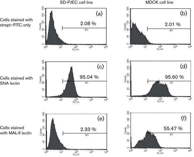
Fig. 2.
Influenza virus receptor expression in SD-PJEC and MDCK cell lines. Both SD-PJEC and MDCK cell lines were incubated with biotinylated MAL-II and SNA lectins followed by staining with streptavidin–FITC. The negative control cells for both cell lines were stained with streptavidin–FITC only. Stained samples were subjected to flow cytometric analysis. A representative experiment out of four experiments is shown here.

