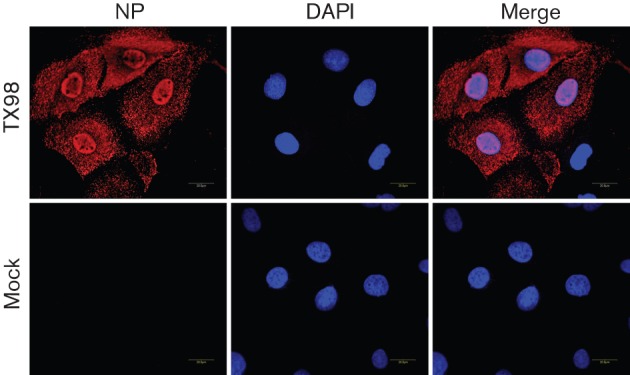
Fig. 3.

Immunofluorescence microscopy detection of NP expression in SD-PJEC-infected cells by A/swine/Texas/4199-2/98 (TX98). Confluent cells were infected with the influenza virus at an m.o.i. of 0.01. At 24 h p.i., cells were fixed and stained with a primary mAb to NP and Alexa Fluor 546-labelled goat anti-mouse antibody was used as secondary antibody. The nucleus was stained with DAPI. Mock-infected cells were used as a control. Specimens were visualized on a Zeiss LSM510 confocal microscope. A 0.8 mm slice through the nucleus is shown in each image.
