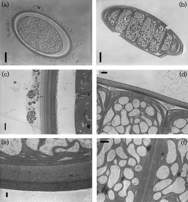
Fig. 3.

Microphotographs of cyanobacterial filaments obtained by transmission electron microscopy. Filament transections of Moorea producens 3LT (a) and M. producens JHB (b); polysaccharide sheaths and thylakoid arrangements in M. producens 3LT with heterotrophic bacteria on the exterior (c), M. producens JHB (d), polysaccharide sheath of M. producens 3LT (e) and thylakoid arrangements in adjacent cells in M. producens JHB (f). Bars: a, 10 µm; b, 10 µm; c, 1 µm; d, 2 µm; e, 0.5 µm; f, 2 µm.
