
Fig. 4.
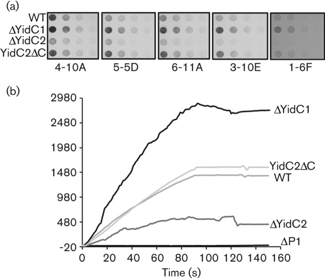
Anti-P1 mAb reactivity with wild-type (WT) and yidC mutant whole cells and comparison of bacterial binding to an agglutinin-coated chip surface. (a) Whole cell dot-blot. Whole bacterial cells serially diluted twofold beginning at 5×10⁶ c.f.u. were applied to nitrocellulose membranes and reacted with the anti-P1 mAb indicated below each blot. (b) Surface plasmon resonance (BIAcore) was used to measure adherence of wild-type (WT) and ΔYidC and ΔP1 mutant cells to a salivary agglutinin-coated chip surface. The sensogram shows the change in resonance units following injection of a suspension of the indicated S. mutans background.