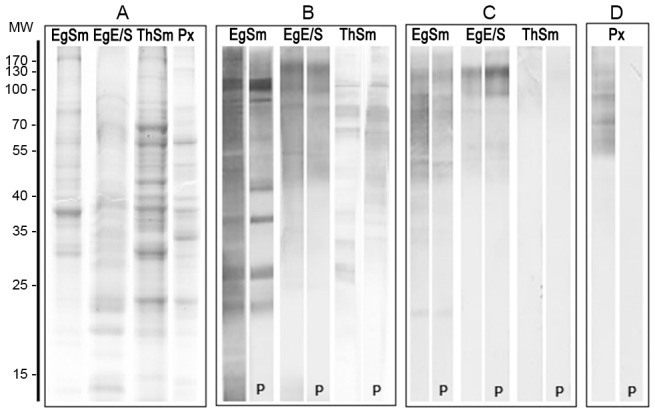
Figure 1. Analysis of parasite antigens by SDS-PAGE and immunoblot.

A) Coomassie stained 12% SDS-PAGE of E. granulosus antigens: E/S, excretion/secretion antigen; Sm, somatic antigen; Px, protoescoleces sonicate; and T. hydatigena somatic antigens: ThSm. Molecular markers in kDa are shown on the left. B–D) Immunoblot probed with polyclonal rabbit antibody PAbCGB10 (B), MAbEg9 (C) or E492/G1 (D). The nitrocellulose strips marked with a “P” were pre-treated with periodate to modify the sugar epitopes.
