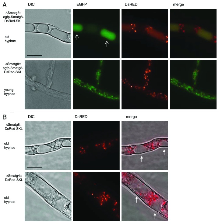
Figure 9.Smatg8 and Smatg4 are involved in pexophagy. (A) Localization of EGFP-SmATG8 and DsRed-SKL in basal hyphae (upper panel) and in apical hyphae (lower panel) of ∆Smatg8. Arrows indicate vacuoles. In basal hyphae DsRed-SKL and EGFP-SmATG8 are localized to the lumen of vacuoles. In apical hyphae peroxisomes are not degraded in the vacuoles. (B) DsRed-SKL is excluded from the vacuole in basal hyphae of ∆Smatg8 and ∆Smatg4. Arrows indicate vacuoles free of the DsRed-SKL fluorescence signals or cellophane. Scale bar: 10 µm.

An official website of the United States government
Here's how you know
Official websites use .gov
A
.gov website belongs to an official
government organization in the United States.
Secure .gov websites use HTTPS
A lock (
) or https:// means you've safely
connected to the .gov website. Share sensitive
information only on official, secure websites.
