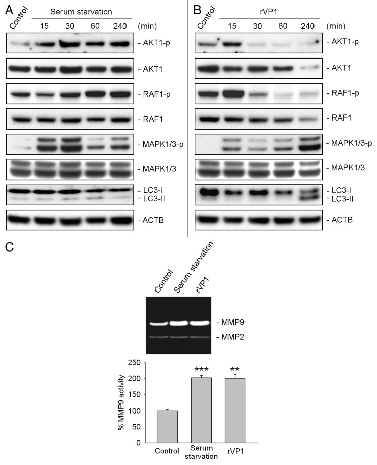
Figure 6. rVP1 increased phosphorylation of MAPK1/3 and MMP9 activity. RAW 264.7 cells were treated with serum starvation or 4 μM rVP1 for 15 to 240 min as indicated. (A and B) Cell lysates were collected and subjected to immunoblot analysis using antibodies against LC3, phosphorylated AKT1 at Ser473, phosphorylated RAF1 at Ser338, phosphorylated MAPK1 (Thr185/Tyr187), phosphorylated MAPK3 (Thr202/Tyr204) and their non-phosphorylation control. ACTB was used as a loading control. Blots are representative of three independent experiments. (C) RAW 264.7 cells were treated with serum starvation or 4 μM rVP1 for 24 h and cell conditioned media were collected. MMP activity was examined by gelatin zymographic analysis. Data represent means ± SEM of three independent experiments; **p < 0.01, ***p < 0.001.

An official website of the United States government
Here's how you know
Official websites use .gov
A
.gov website belongs to an official
government organization in the United States.
Secure .gov websites use HTTPS
A lock (
) or https:// means you've safely
connected to the .gov website. Share sensitive
information only on official, secure websites.
