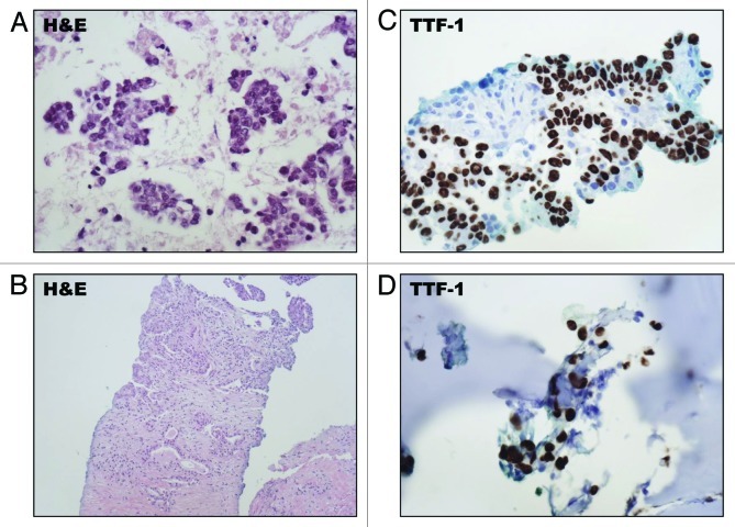
Figure 2. Histopathologic features of the EML4-ALK translocation positive tumor from patient 1. (A) An FNA of the patients left lung tumor showed malignant cells with hematoxylin and eosin (H&E) staining. (B) A core needle biopsy of the primary left lower lobe lung tumor showed moderately-differentiated adenocarcinoma. (C) The tumor cells exhibited nuclear expression of TTF-1 by IHC. (D) A biopsy of a nasal bone lesion confirmed metastatic disease which similarly expressed TTF-1 by IHC.

An official website of the United States government
Here's how you know
Official websites use .gov
A
.gov website belongs to an official
government organization in the United States.
Secure .gov websites use HTTPS
A lock (
) or https:// means you've safely
connected to the .gov website. Share sensitive
information only on official, secure websites.
