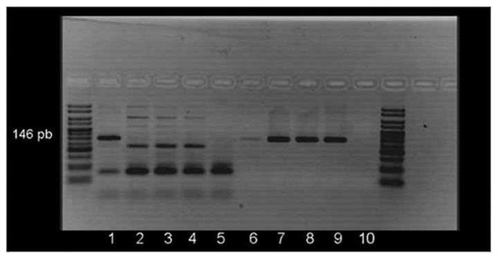
Figure 2. Methylated and unmethylated RARβ PCR amplification of two patients with malignant pleural effusion. Lanes 1–5 correspond to the methylated-specific PCR and lanes 6–10 to the unmethylated-specific PCR. Lanes 1 and 6: methylated control; 2 and 7: unmethylated control; 3 and 4: two samples (no 146 pb band appears, indicating no methylation); 8 and 9: the same samples (a 146 pb appears indicating no methylation); 5 and 10: negative control.

An official website of the United States government
Here's how you know
Official websites use .gov
A
.gov website belongs to an official
government organization in the United States.
Secure .gov websites use HTTPS
A lock (
) or https:// means you've safely
connected to the .gov website. Share sensitive
information only on official, secure websites.
