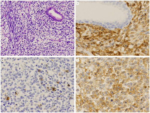
Figure 3.

Microscopic findings. a)The tumor was made up of sarcomatoid oval to spindle cells. (HE x 100) b) Immunostaining with CD56 showed positive results for tumor cells on the cell membrane (CD56 x 200). c) Immunostaining with synaptophysin focally showed positive results for the tumor cells in the cytoplasm (synaptophysin x 200). d) Immunostaining with CD99 showed positive results for tumor cells on the cell membrane (CD99 x 200).
