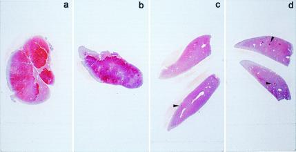
Figure 1.
Immunohistochemical staining for GST 7-7 (red) of liver sections from rats killed at 2 months after Tx. (a and b) Group IA: 80–90% of the liver was positive for GST 7-7. (c) Group IIA: Small positive foci are present (arrowhead). (d) group IB: A few GST 7-7-positive foci were detected (arrowheads); see text for details.

