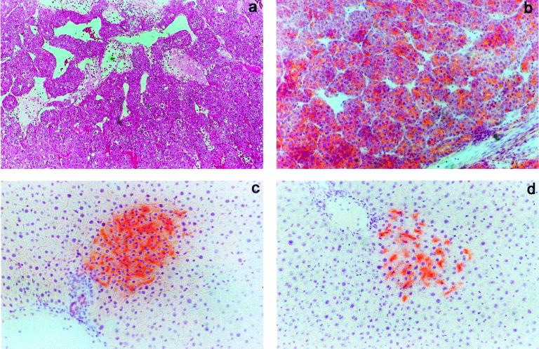
Figure 3.
(a and b) Liver samples from a rat in group IA killed 4 months after Tx. (a) H&E-stained section showing a well differentiated hepatocellular carcinoma with areas of necrosis. (b) Histochemical staining for DPPIV (orange) documenting that the cancer cell population was positive for the enzyme, i.e., it was from donor origin. (c) Liver sample from a rat in group IIA killed 6 months after Tx; the animal underwent two-thirds PH 2 months before killing. Histochemical staining for DPPIV (orange) revealed that donor-derived hepatocyte clusters were still present in these animals; however, their growth was very limited. (d) Liver sample from a rat in group IIB killed 6 months after Tx; the animal underwent two-thirds PH 2 months before killing. Staining for DPPIV enzyme activity documented the persistence of donor-derived cell clusters. (Original magnification ×100.)

