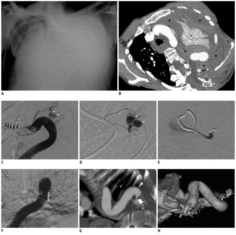
Fig. 1.
Spontaneous massive hemothorax in 46-year-old woman with neurofibromatosis type 1.
A. Antero-posterior chest film reveals haziness of left lung, mediastinum shift to right, and severe kyphoscoliosis. B. CT angiography of thoracic aorta shows small aneurysm (arrow) with massive blood clot (asterisks) associated with total left lung atelectasis. (C) Aortography and (D) selective left 5th intercostal arteriography demonstrate saccular aneurysm (small arrow) at proximal left 5th intercostal artery with dilated adjacent vein (arrow head) due to arteriovenous fistula between left 5th intercostal artery and vein, which drains into hemiazygose vein (multiple arrows) (E) Left 5th intercostal arteriography after coil embolization shows complete occlusion of pseudoaneurysm and arteriovenous fistula. However, aneurysmally dilated intercostals artery remains even after embolization of pseudoaneurysm.
F. Aortography after embolization shows complete occlusion of pseudoaneurysm and arteriovenous fistula with absence of other potential bleeding. (G) Maximal intensity projection image in coronal viewand (H) volume rendered image of CT angiography at 1 year after coil embolization show complete occlusion of prior pseudoaneurysm. However, there is recurrence of small arteriovenous fistula between intercostal artery (thin arrow) and vein (arrow head). Note metallic coil (thick arrow).

