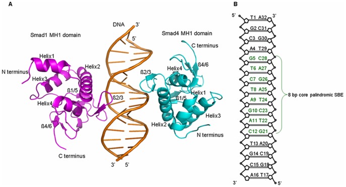
Figure 1. Structure of Smad1 MH1 and Smad4 MH1 bound to DNA and.
Sketch of DNA. (A) The overall structure of Smad1 MH1 and Smad4 MH1 domains bound to SBE palindrome DNA shown in cartoon form with Smad1 MH1 colored in magenta on the left and Smad4 MH1 colored in cyan on the right; (B) Schematic drawing of the 16-bp palindrome DNA sequence with the eight core base pairs colored in dark-green.

