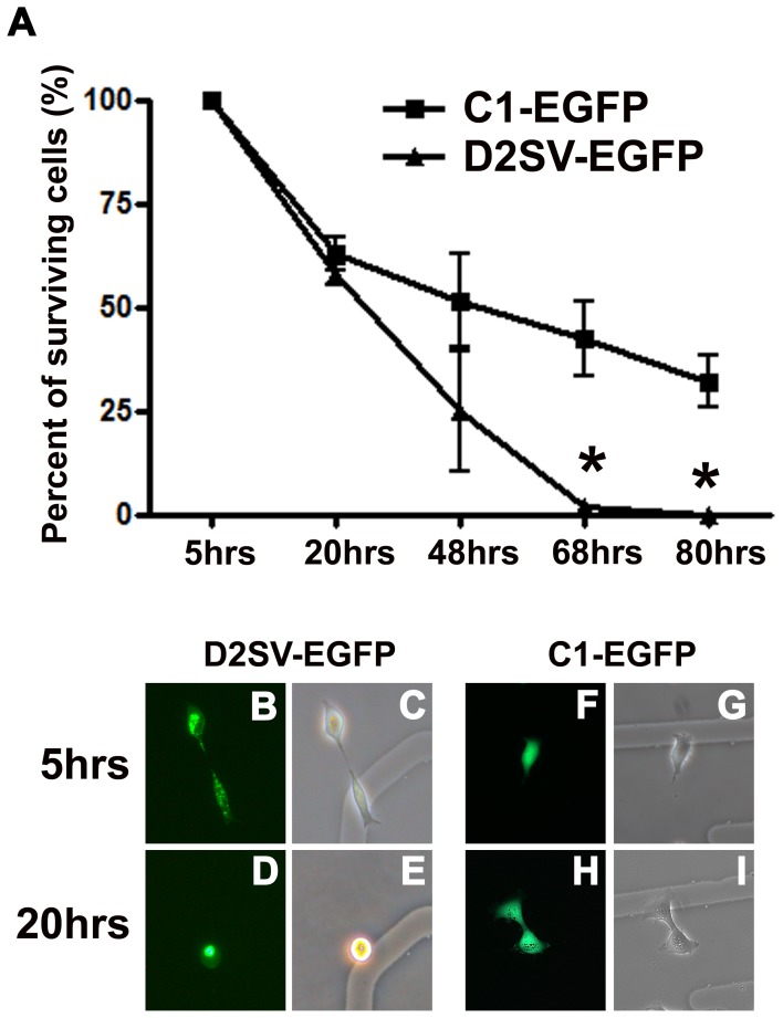
Figure 9. Analysis of D2SV-EGFP induced cell death in collaboration with trypsinization.
HEK293 cells transfected with D2SV-EGFP or C1-EGFP were trypsinized and reseeded on gridded coverslips. In each experiment, cells were followed for 80 hrs to determine long term cell viability after trypsinization. Surviving cells were quantified and expressed as a percent of total counted cells (A). Values are expressed as mean ± SEM. One-way ANOVA, *p<0.05, seven to twenty cells were followed from three independent experiments (N = 3). Photomicrographs depict examples of D2SV-EGFP (B–E) and C1-EGFP (F–I) cells at 5 hrs (B, C, F, G) and 20 hrs (D, E, H, I). Note all EGFP-D2SV transfected cells disappeared from culture by 68 hrs post trypsinization (A). Examples of cell death in EGFP-D2SV transfected cells as observed in live cultures by fluorescence microscopy (B–E).

