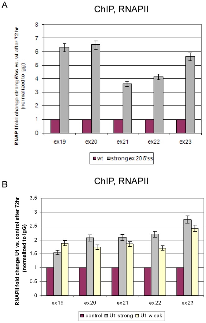
Figure 4. Splicing affects RNAPII occupancy.

(A) At 72 hr following transfection with either the wt or strong exon 20 5′ss minigene, cells were collected and used for an RNAPII-ChIP analysis. The precipitated DNA fragments were subjected to QPCR. Enrichment values were normalized to the unbound fraction, to a non-specific IgG antibody, and to the GFP area of the plasmid. Results are presented as RNAPII fold change between strong exon 20 5′ss and the wt plasmid. (B) At 72 h following co-transfection of IKAP19–23 and U1 plasmids, RNAPII-ChIP was performed. All experiments were repeated independently three times, and the results shown are representative of an average experiment. QPCR experiments were amplified in triplicate; results shown are mean values ± SD.
