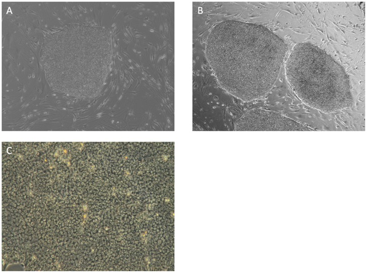
Figure 1. Morphological characteristics of hiPSCs.
Typical phase contrast pictures of UCBiP7 (A) and PCBC16ShiP (B) grew in flat and compact colonies with distinct cell borders in monolayer culture with irradiated MEF (Magnification = 40x). (C) Microscopic 100× view of a colony of undifferentiated hiPSC: high nucleus-to-cytoplasm ratios, and prominent nucleoli.

