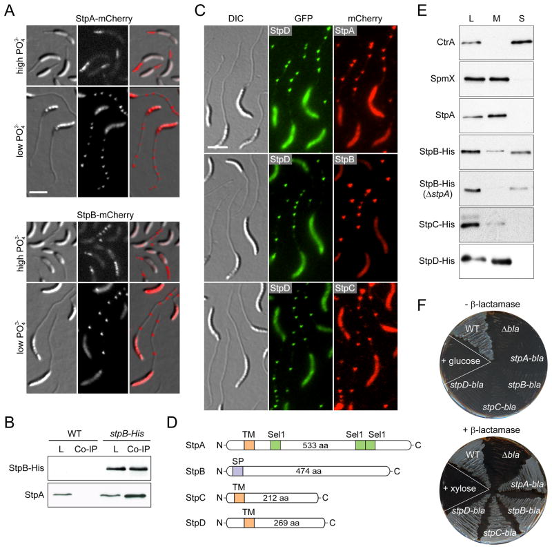
Figure 2. Identification and subcellular localization of novel stalk proteins.
(A) Stalk localization of StpA-mCherry (SW33) and StpB-mCherry (SW30) produced from the xylose-inducible Pxyl promoter after 24 h of growth in phosphate-rich (M2G, high PO43−) or phosphate-poor medium (M2G−P, low PO43−) containing 0.3% xylose.
(B) Co-immunoprecipitation analysis of stpB-His (SS233) and wild-type cells reveals an interaction between StpA and StpB. Whole-cell lysates (L) and eluates from co-immunoprecipitation experiments (Co-IP) were subjected to immunoblot analysis using anti-His and anti-StpA antibodies.
(C) StpA,B,C,D localization in stalks is reminiscent of the distribution of crossbands. Cells of strain SS243 (stpD::stpD-gfp Pxyl::Pxyl-stpA-mcherry), SS88 (stpB::stpB-mcherry stpD::stpD-gfp) and SS389 (stpC::stpC-mcherry stpD::stpD-gfp) were grown in M2G−P for 24 h. Synthesis of StpA-mCherrry was induced with 0.3% xylose for 24 h.
(D) Schematic depicting the domain organization of the stalk proteins with the predicted transmembrane domains (orange), the signal peptide (purple) and the Sel1 motifs (green).
(E) Cell fractionation analysis reveals that StpA, StpC, and StpD are membrane-bound proteins, whereas StpB is soluble. Whole-cell lysates (L) and the corresponding membrane (M) and soluble (S) fractions of cells producing His-tagged Stp proteins (SS233, SS220, SS244 and SS247) were subjected to Western blot analysis using an anti-His antibody. Fractionation efficiency was verified by probing the same fractions with anti-CtrA and anti-SpmX antibodies. Note, the absence of stpA and stpAB does not reduce the levels of StpB and StpC, respectively (Figure S2C).
(F) The Stp proteins are targeted to the periplasm. The TEM-1 β-lactamase gene (bla) was fused to the 3’ end of stpA, stpB, stpC and stpD, respectively. The gene fusions were placed under the Pxyl promoter in a β-lactam-sensitive reporter strain. Cells (SS165, SS172, SS273, SS274) were patched on PYE agar containing ampicillin and either 0.2% glucose or 0.3% xylose. Scale bars: 3 μm. See also Figure S2.

