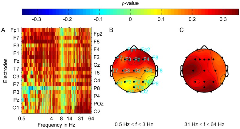
Figure 2. Spatio-spectral distribution of correlation strength between tinnitus loudness and oscillatory band power for the subgroup with pure tone tinnitus.
Group averages are shown. Power spectra were interpolated with a resolution of 40 points per 1 Hz. Tinnitus loudness was determined by adjusting the contribution of each frequency component and the loudness of such a reconstructed tinnitus spectrum to the perceived tinnitus. Correlations were controlled for age, global psychological distress (GPD), and mean hearing loss (MHL) between 0.125 kHz and 16 kHz. (A) Correlation strength (Spearman's  ) at each electrode and frequency point is shown. Plots (B) and (C) show correlation maps corresponding to (A) with averaged correlation strength (
 ) topographies for the tinnitus loudness
  and delta (B) or gamma (C) oscillatory power. Correlation strength for delta band power and tinnitus loudness was highest in the frontal half of the brain and lowest at posterior locations. For the correlation between gamma band power and tinnitus loudness the distribution of correlation strength across electrode positions was more uniform. Highest correlation strength was reached at the left temporal and right occipital electrode positions. After FDR correction (FDR 0.05:
  ) correlations remained significant at all electrode positions except for T8 and P8 locations for the gamma band, whereas significant correlations in the delta band were attained at the fronto-central locations Fp2, F1, Fz, F2, F4, F8, C3, Cz, and at P7.

