Fig. 2.
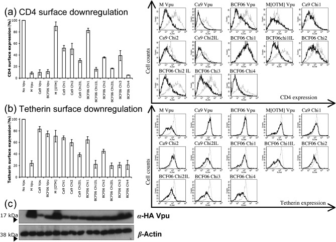
M and O group Vpus downregulate CD4 and tetherin differently from the cell surface. (a) 293T cells were cotransfected with a CD4 expression vector and pIRES2eGFP plasmids expressing GFP alone (no Vpu) or together with the indicated Vpu constructs, and CD4 surface expression was measured by FACS. Shown are relative levels of CD4 cell surface expression (black line) relative to those measured in cells transfected with the GFP only control vector (dotted line). (b) 293T cells were cotransfected with an ectodomain HA-tagged human tetherin vector and pIRES2eGFP plasmids expressing GFP alone (no Vpu) or together with the indicated Vpu constructs and HA tetherin surface expression was measured by FACS. Shown are the relative levels of tetherin cell surface expression (black line) relative to those measured in cells transfected with the GFP only control vector (dotted line). Results are mean±sem of three separate experiments. (c) Western blot of HA-tagged Vpu in the pIRES contruct showing expression of the various Vpu constructs, as well as a β-actin control.

