Figure 1.
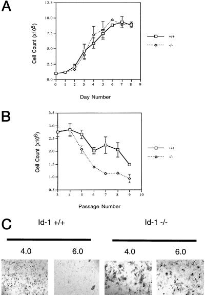
Id1 −/− MEFs exhibit premature cellular senescence. (A) Growth curves for Id-1 +/+ and −/− MEFs at P-3. Replicate cultures of 1 × 10⁵ cells per 35-mm diameter dish were plated on day zero and cells were counted each day for 8 consecutive days (diao). (B) Senescence profile of Id-1 +/+ and −/− MEFs. P-3 MEFs were passaged on a 3T9 protocol in which 9 × 10⁵ cells were plated in a 60-mm dish and counted every 3 days with subsequent replating of 9 × 10⁵ cells at each passage until senescence was reached. (C) β-gal activity of P-8 MEFs. Cells were stained at pH 4.0 for lysosomal β-gal activity (positive control) and pH 6.0 for senescence-associated β-gal activity. Positive control is included to detail cellular morphology of Id1 +/+ vs. −/− cells at P-8 (×100).

