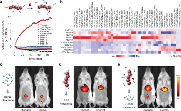
Figure 2. Urinary detection of in vivo protease activity with peptide-NWs.
(a) Shown are representative activation profiles of peptide-NWs following treatment with recombinant proteases. Specific protease-substrate combinations led to rapid increases in sample fluorescence. (b) Heat map comparison of cleavage velocities for different substrate-protease combinations grouped according to activity and specificity. (c) Fluorescence in vivo images of DDC-treated and control animals following intravenous injection of VivoTag-680 labeled Glu-fib peptides, (d) peptide-free NWs or (e) peptide-conjugated NWs.

