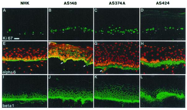
Figure 3.
Increased proliferation in XP-C epidermis reconstructed in vitro on a normal dermal equivalent. (A, E, and I) Normal epidermis reconstructed from normal human keratinocytes (NHK). (B–D, F–H, J–L) XP-C epidermis reconstructed from XP-C keratinocyte strains AS148, AS374A, and AS424, as indicated. Immunolabeling: A–D, proliferation-associated Ki-67 antigen (Ki 67); E–H, α-6 integrin; I–L, β-1 integrin (beta 1). Note that virtually all nuclei are labeled with anti-Ki-67 antibody in all XP-C epidermis, whereas about 10% only are found in normal epidermis (NHK). Also note the increased deposition of α-6 and β-1 integrins at the basement membrane zone of XP-C compared with normal epidermis. In XP-C epidermis also, β-1 integrin is abnormally expressed in several suprabasal layers above the basal layer, whereas it is restricted in normal epidermis. Arrows point to the nascent epidermal invasions within the dermal equivalent, as indicated by β-1 integrin staining. The dashed line indicates the dermal–epidermal junction. (Bar = 50 μm.)

