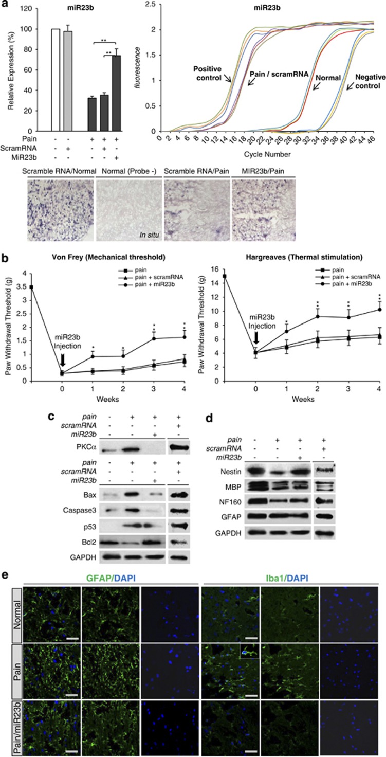
Figure 1.
The intrathecal administration of miR23b reduces neuropathic pain in an animal model. (a) The expression level of miR23b was significantly increased after intrathecal injection of miR23b in a neuropathic pain model, as determined by RT-PCR (**P<0.01) and in situ hybridization (ISH) analysis (bottom). (b) The PWT was determined using both von Frey filaments to measure the mechanical threshold and the Hargreaves method to measure noxious thermal sensitivity. After the injection of miR23b or scramRNA, mechanical and thermal hypersensitivity were attenuated for 4 weeks. (*P<0.05; **P<0.01). (c) Western blot analyses were used to determine the expression levels of survival- and death-related genes. Intrathecal administration of miR23b-induced STAT3 expression and Akt activation, whereas the expression of the apoptotic cell death signals p38, JNK, Bax, and caspase-3 was reduced. (d) Furthermore, the neural markers nestin, MBP, and neurofilament 160 (NF160) were increased after intrathecal administration of miR23b. (e) The astrocyte cell marker glia fibrillary acidic protein (GFAP) and the microglia cell marker Iba1 were increased following induction of neuropathic pain, whereas injured mice receiving miR23b infusions showed decreased expression of these markers. GFAP and Iba1 were immunolabeled with anti-GFAP and anti-Iba1 antibodies, respectively

