Figure 6.
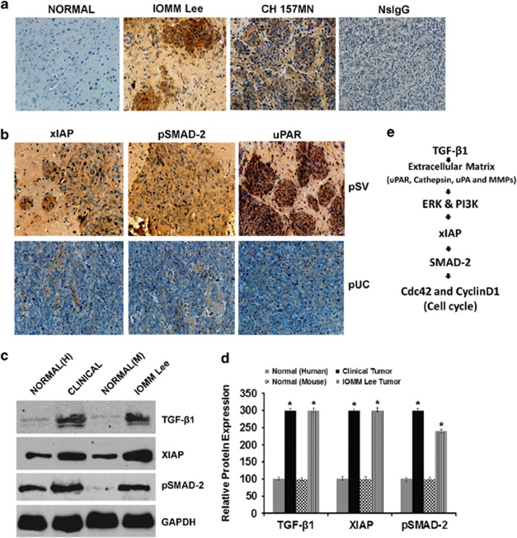
Knockdown of uPAR and cathepsin B reduces TGF-β1-induced signaling in vivo. (a) Nude mice were intracranially implanted with IOMM-Lee and CH157-MN cells as described in Materials and Methods. Formalin-fixed brain sections were subjected to immunostaining with anti-TGF-β1 antibody and nonspecific IgG (NsIgG). Representative photomicrographs ( × 200) are shown in comparison with normal mouse brain sections. (b) Nude mice intracranially implanted with IOMM-Lee cells were subsequently treated with pSV and pUC in different groups as described earlier. Formalin-fixed brain sections were checked for immunoreactivity with XIAP, pSMAD-2, and uPAR antibodies, and representative photomicrographs ( × 200) are shown. (c) Frozen brain tissue lysates either from animal models or clinical samples were blot transferred and analyzed for TGF-β1, XIAP, and pSMAD-2; GAPDH served as a loading control (H, human; M, mouse). (d) The band intensities from each blot were quantified with ImageJ software and relative expression levels were plotted. Each column designates mean±S.D. of three independent experiments. *P<0.05, significant difference from respective normal tissues. (e) Schematic representation of TGF-β1 induced signaling in meningioma cells

